


Why Do Pyogenic Granulomas Bleed So Easily?


Pyogenic granulomas bleed so easily because they are made up of rapidly growing, abnormally fragile blood vessels packed just beneath a thin layer of skin. At Hazany Derm, Dr. Salar Hazany provides precise, minimally invasive removal to stop recurrent bleeding and restore healthy skin.
If you have ever nicked a pyogenic granuloma while shaving, bumped it against a towel, or simply touched it too firmly, you already know the alarming result: bright red blood that seems wildly out of proportion to such a small skin growth. This reaction surprises most patients, who often wonder whether something more serious is happening. The truth lies in the very nature of what a pyogenic granuloma actually is at a structural level.
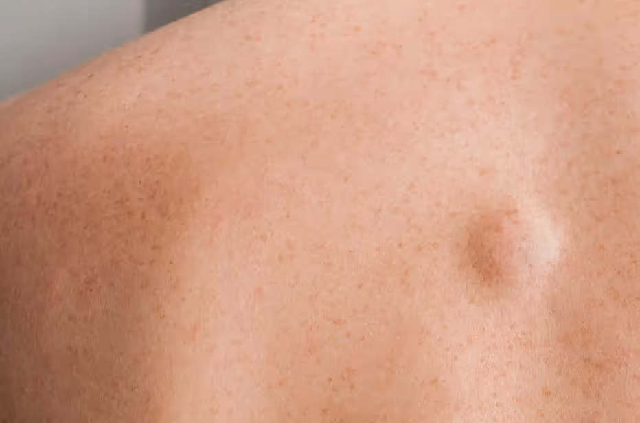
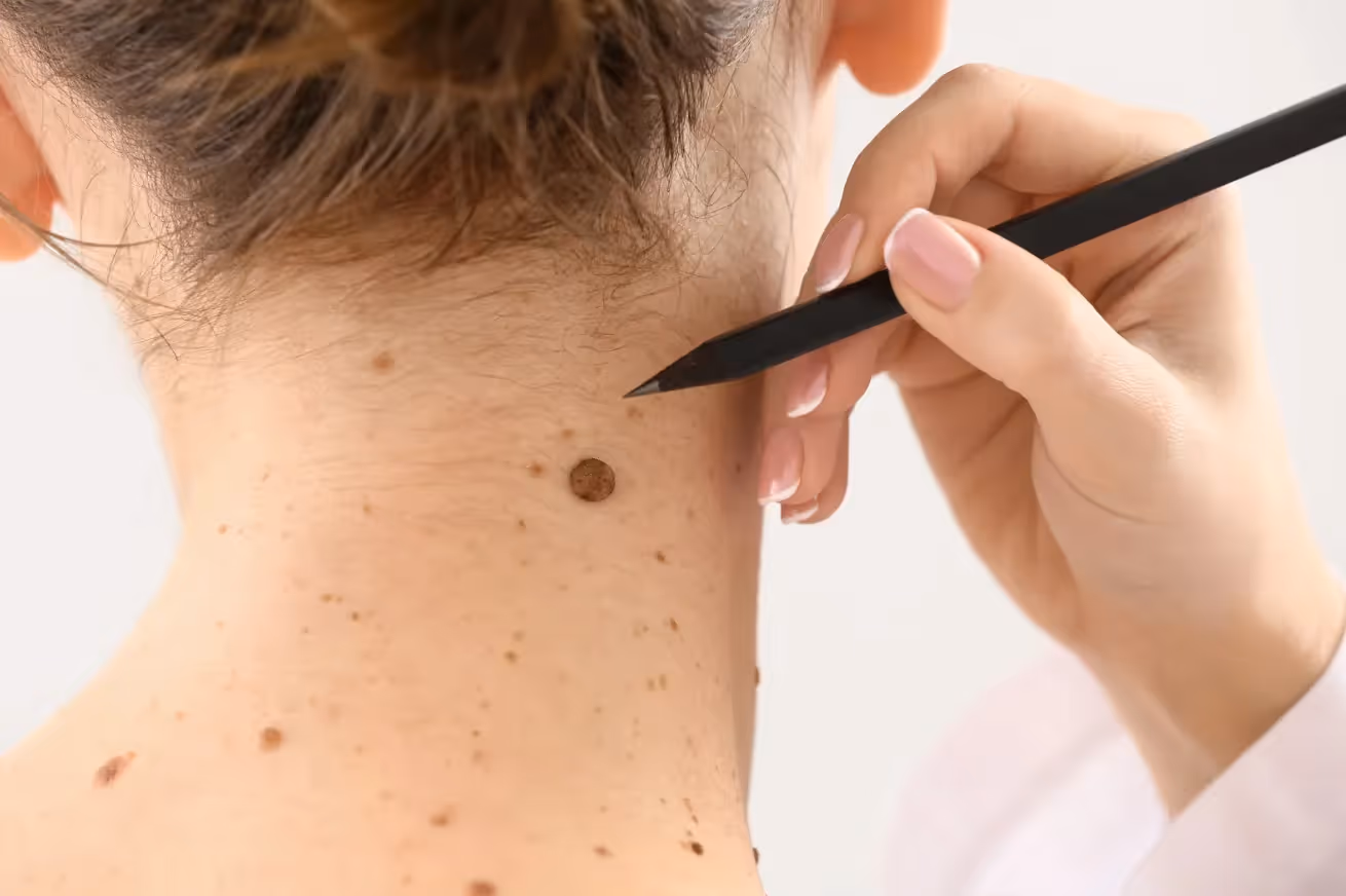
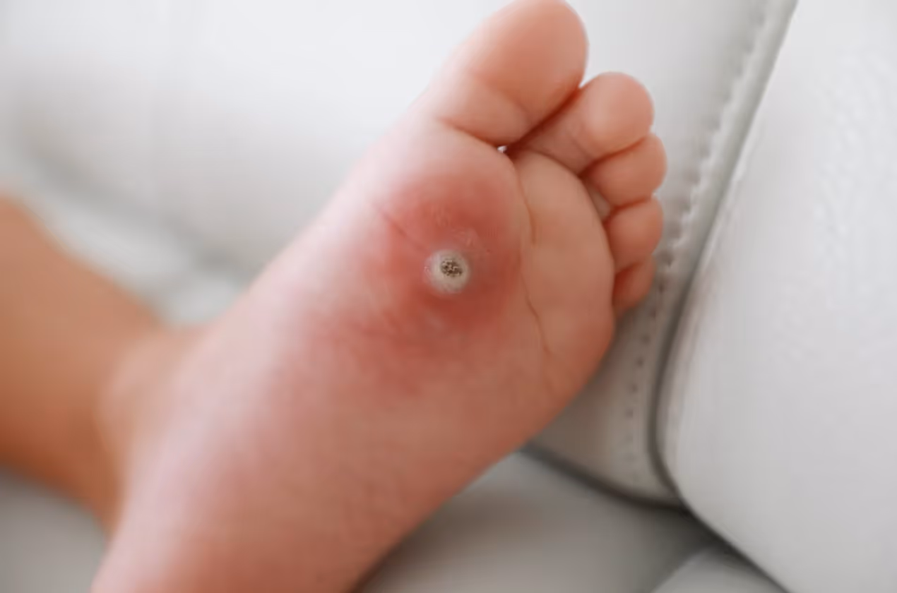
Pyogenic granulomas are small, raised, red or reddish-brown bumps that appear on the skin or mucous membranes. They are not cancerous, and despite their alarming name, they are not caused by bacteria or pus as the term "pyogenic" might suggest. They are, in fact, benign overgrowths of blood vessel tissue that tend to grow rapidly over a period of days to weeks. This rapid, disorganized growth is precisely what makes them so prone to bleeding.
Understanding why these growths bleed the way they do helps explain why proper treatment is so important. Leaving a pyogenic granuloma untreated is not simply an aesthetic choice - it can mean dealing with repeated, sometimes heavy bleeding episodes that interfere with daily life. At Hazany Derm in Los Angeles, Dr. Salar Hazany evaluates and removes these lesions with the precision and care needed to address both the cosmetic concern and the functional problem of recurrent bleeding.
The Vascular Architecture of a Pyogenic Granuloma
To understand why pyogenic granulomas bleed so readily, you first need to understand what they are made of. At their core, these lesions are a type of benign vascular tumor, meaning they are composed almost entirely of blood vessel tissue. The technical term used in dermatology and pathology is lobular capillary hemangioma, which tells you everything about their structure: lobules, or clusters, of tiny capillaries packed densely together within a small area of skin.
The capillaries inside a pyogenic granuloma are not ordinary, mature blood vessels. They form through a process called angiogenesis, which is the rapid growth of new blood vessels, often triggered by minor trauma, hormonal changes, or skin irritation. This new vessel growth happens quickly and without the normal architectural support that healthy tissue develops over time. The result is a tangle of immature, thin-walled capillaries that are structurally fragile and extremely close to the skin surface.
What makes these vessels particularly vulnerable is the combination of high density and poor structural support. In normal skin, blood vessels are surrounded by connective tissue that stabilizes them and reduces the risk of rupture from minor contact. In a pyogenic granuloma, the surrounding stroma is loose, edematous, and poorly organized. This means the vessels have almost no protective scaffolding around them, and even the gentlest mechanical force can cause them to rupture and bleed profusely.
The four structural reasons pyogenic granulomas bleed so easily include:
- Thin vessel walls: The capillaries within the lesion are immature and lack the muscular reinforcement found in fully developed blood vessels, making them extremely easy to rupture.
- Superficial location: Pyogenic granulomas sit very close to the outermost layer of skin, meaning the vessels have almost no overlying tissue to protect them from external contact.
- High capillary density: The sheer number of blood vessels packed into a small area means that even a tiny wound disrupts multiple vessels simultaneously, producing bleeding that seems disproportionate to the size of the injury.
- Lack of fibrous support: The connective tissue surrounding the vessels is loose and poorly formed, offering little structural resistance when pressure or friction is applied.
These structural characteristics make pyogenic granulomas behave very differently from a simple cut or abrasion. A normal skin wound bleeds because a vessel is severed and then clotting mechanisms take over relatively quickly. With a pyogenic granuloma, the vessel walls are so thin and the density is so high that bleeding can restart repeatedly even after it appears to have stopped. Patients often describe pressing firmly on the lesion, watching the bleeding slow, releasing pressure, and then seeing it start again within seconds.
This is why Dr. Salar Hazany at Hazany Derm emphasizes that watchful waiting is rarely the right approach for these lesions once they begin to cause bleeding problems. The structural fragility does not resolve on its own, and the lesion does not simply remodel into stronger, more stable tissue over time. Definitive removal is the most reliable way to stop the bleeding cycle entirely.
Why Certain Locations Bleed More Than Others
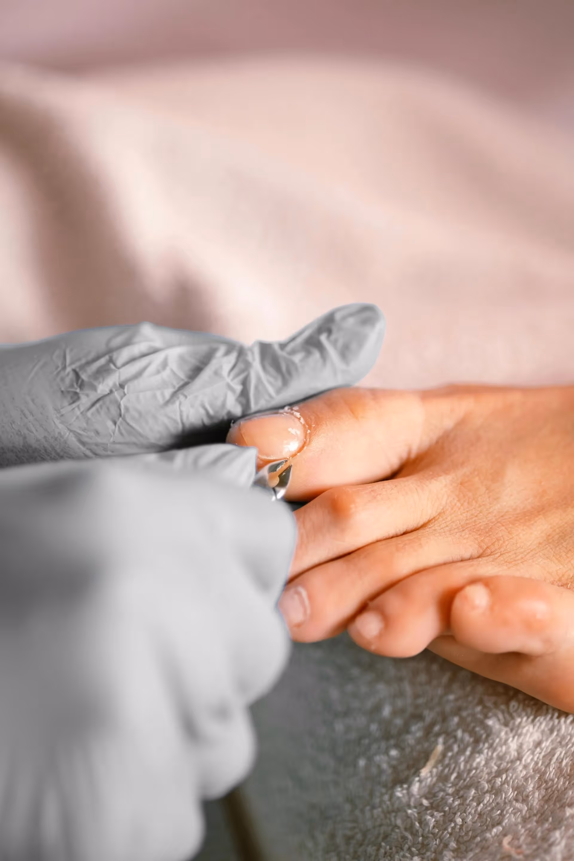
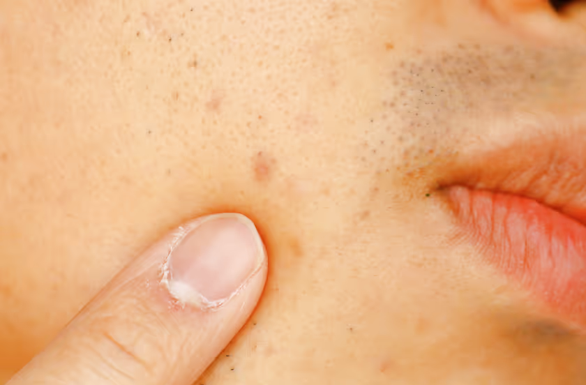
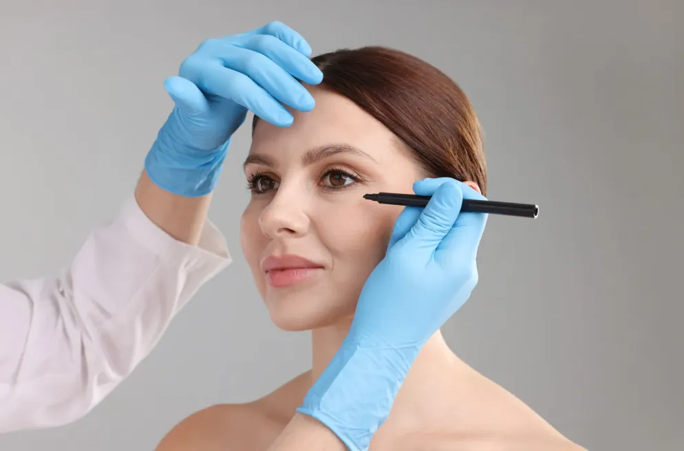
The location of a pyogenic granuloma on the body has a significant influence on how severely and how often it bleeds. Lesions that form on areas exposed to repeated friction, movement, or accidental contact tend to bleed far more frequently than those in sheltered locations. Some of the most problematic sites include the face, particularly the nose, lip, and eyelid, as well as the fingers and hands where contact with surfaces is constant.
The nose is one of the most common and most troublesome sites for pyogenic granulomas. Nasal skin is thin, the underlying tissue is richly vascularized, and even gentle pressure from glasses frames, makeup application, or blowing the nose can be enough to trigger bleeding. A pyogenic granuloma on the nose can look concerning because of its bright red, dome-shaped appearance, and it bleeds easily enough to cause staining on clothing and real anxiety for the patient. Dr. Salar Hazany routinely addresses these facial lesions at Hazany Derm using techniques designed to minimize scarring while achieving complete removal.
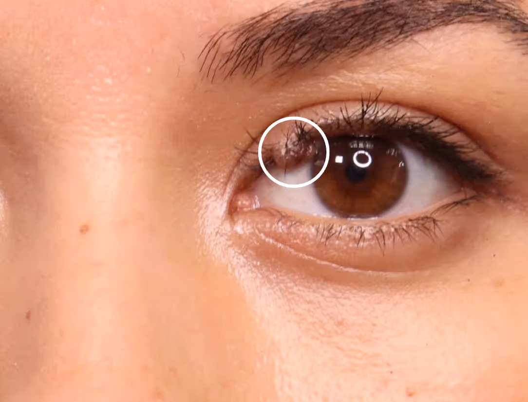
The lip and eyelid present their own set of challenges. The lip is constantly in motion during speaking, eating, and drinking, which means a lesion in that area faces repeated mechanical stress throughout every single day. The eyelid is similarly active and extremely delicate, with skin so thin that the underlying vascularity is essentially right at the surface. Growths in these areas are particularly prone to spontaneous bleeding even without direct trauma, simply because of the constant movement and the minimal tissue buffer between the lesion and the external environment.
The four location-related factors that influence bleeding frequency and severity are:
- Friction exposure: Areas of the skin that experience regular contact with clothing, jewelry, shaving tools, or other surfaces create repeated micro-traumas that rupture the fragile vessels within the lesion.
- Tissue thickness: Sites where the overlying skin is naturally thin, such as the eyelid, lip, and nasal tip, offer far less protection to the underlying capillaries than areas with thicker dermis.
- Movement and muscle activity: Locations near active muscles, such as around the mouth or on the fingers, experience constant mechanical stress that prevents the lesion from remaining undisturbed long enough to stop bleeding naturally.
- Vascularity of the region: Some areas of the body are inherently more richly supplied with blood vessels, meaning that lesions in those regions have more vascular input and bleed more heavily when disrupted.
The combination of an already fragile lesion structure and a high-friction, high-movement location creates conditions where bleeding becomes almost unavoidable on a daily or weekly basis. Patients with lesions on the lip often report bleeding while eating or brushing their teeth. Those with finger lesions frequently bleed at work or while performing routine tasks. This persistent disruption to daily life is one of the most compelling reasons patients seek evaluation and treatment.
At Hazany Derm, the approach to skin lesion removal takes into account not just the lesion itself but its precise location and the specific demands placed on that area of the body. Dr. Salar Hazany uses minimally invasive techniques that are particularly well suited to sensitive anatomical sites where scarring and tissue disruption must be kept to an absolute minimum.
What Happens When a Pyogenic Granuloma Bleeds and Why It Keeps Coming Back
One of the most frustrating aspects of living with a pyogenic granuloma is that even when bleeding is controlled with pressure, the lesion almost always bleeds again. Understanding the cycle of bleeding and regrowth helps clarify why home management is limited and why professional removal is typically necessary. The underlying reason is that the lesion itself remains entirely intact even after a bleeding episode, and in some cases the act of bleeding and minor trauma can actually stimulate further growth.
When a pyogenic granuloma bleeds, the body responds with its normal clotting cascade: platelets aggregate, fibrin forms, and a clot develops over the wound surface. However, because the lesion is made up of actively proliferating vascular tissue, the healing response at the wound site can inadvertently signal more blood vessel growth. This is similar to how repeated scratching of certain skin conditions can worsen them over time. The growth factors released during wound healing, particularly vascular endothelial growth factor and other angiogenic signals, can feed the very tissue that caused the problem in the first place.
There is also a physical reason why the lesion bleeds repeatedly even without new growth. Once a pyogenic granuloma has bled and formed a surface crust, that crust is fragile and adheres loosely to the underlying lesion. Daily activities easily dislodge the crust before the surface has truly healed, reopening the wound and exposing the dense capillary network again. Patients sometimes notice that the lesion seems to grow slightly larger after repeated bleeding episodes, and pathologically this can be true - repeated trauma and healing attempts can enlarge the granuloma over weeks to months.
The four key reasons why pyogenic granulomas keep bleeding despite home management include:
- Intact lesion tissue: Pressure and bandaging can stop active bleeding temporarily, but they do nothing to remove the underlying mass of fragile blood vessels that will bleed again as soon as pressure is released and normal activity resumes.
- Wound healing signals: The growth factors released during each healing response can stimulate further capillary proliferation within the lesion, potentially making it larger and more vascular with each bleeding episode.
- Crust fragility: Surface crusts that form over a pyogenic granuloma after bleeding are loosely attached and easily disrupted by movement, washing, or accidental contact before proper wound closure occurs.
- No spontaneous regression in adults: While pyogenic granulomas in certain contexts, such as pregnancy-related lesions, occasionally regress after hormonal changes, lesions in most adults do not resolve on their own and will continue to bleed indefinitely without treatment.
The practical implication of this cycle is that home remedies, silver nitrate sticks, and repeated cautery attempts without complete removal of the base often lead to recurrence. Partial treatment destroys the surface of the lesion but leaves behind the deeper vascular lobules, which can regenerate and produce a new lesion at the same site within weeks. This is one of the most common reasons patients arrive at Hazany Derm after having a pyogenic granuloma treated elsewhere, only to see it return.
Dr. Salar Hazany's approach to pyogenic granuloma removal at Hazany Derm is designed specifically to address this recurrence problem. By removing the lesion thoroughly and addressing the base of the growth, the treatment eliminates the vascular tissue responsible for both the bleeding and the tendency to regrow. Patients treated at Hazany Derm benefit from a careful, precise technique that prioritizes both complete removal and excellent cosmetic outcomes, so they are not left choosing between stopping the bleeding and minimizing the appearance of a scar.
Frequently Asked Questions
Why do pyogenic granulomas bleed so much from such a small bump?
Pyogenic granulomas are essentially dense clusters of immature, thin-walled capillaries packed into a very small area just beneath the skin surface. Because the vessel walls are fragile and the capillary density is extremely high, even minimal contact disrupts multiple vessels at once and produces bleeding that seems disproportionate to the size of the lesion. Dr. Salar Hazany at Hazany Derm explains to patients that this is not a sign of anything dangerous, but it does mean the lesion needs to be properly removed to stop the recurring bleeding cycle.
Is a pyogenic granuloma dangerous if it bleeds heavily?
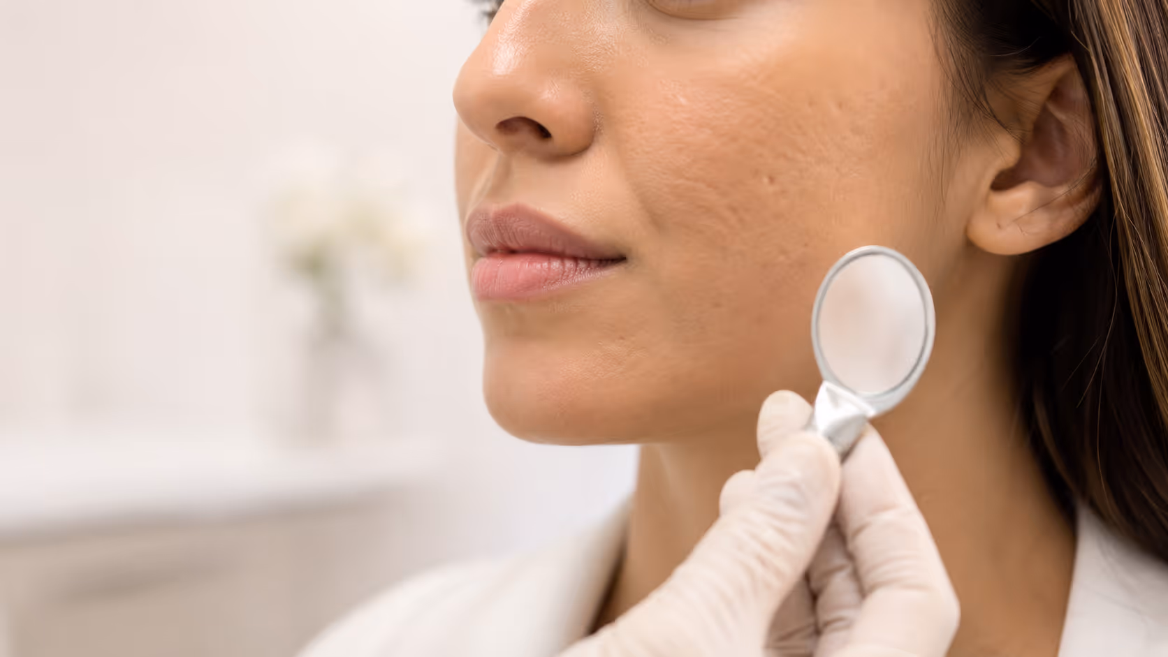
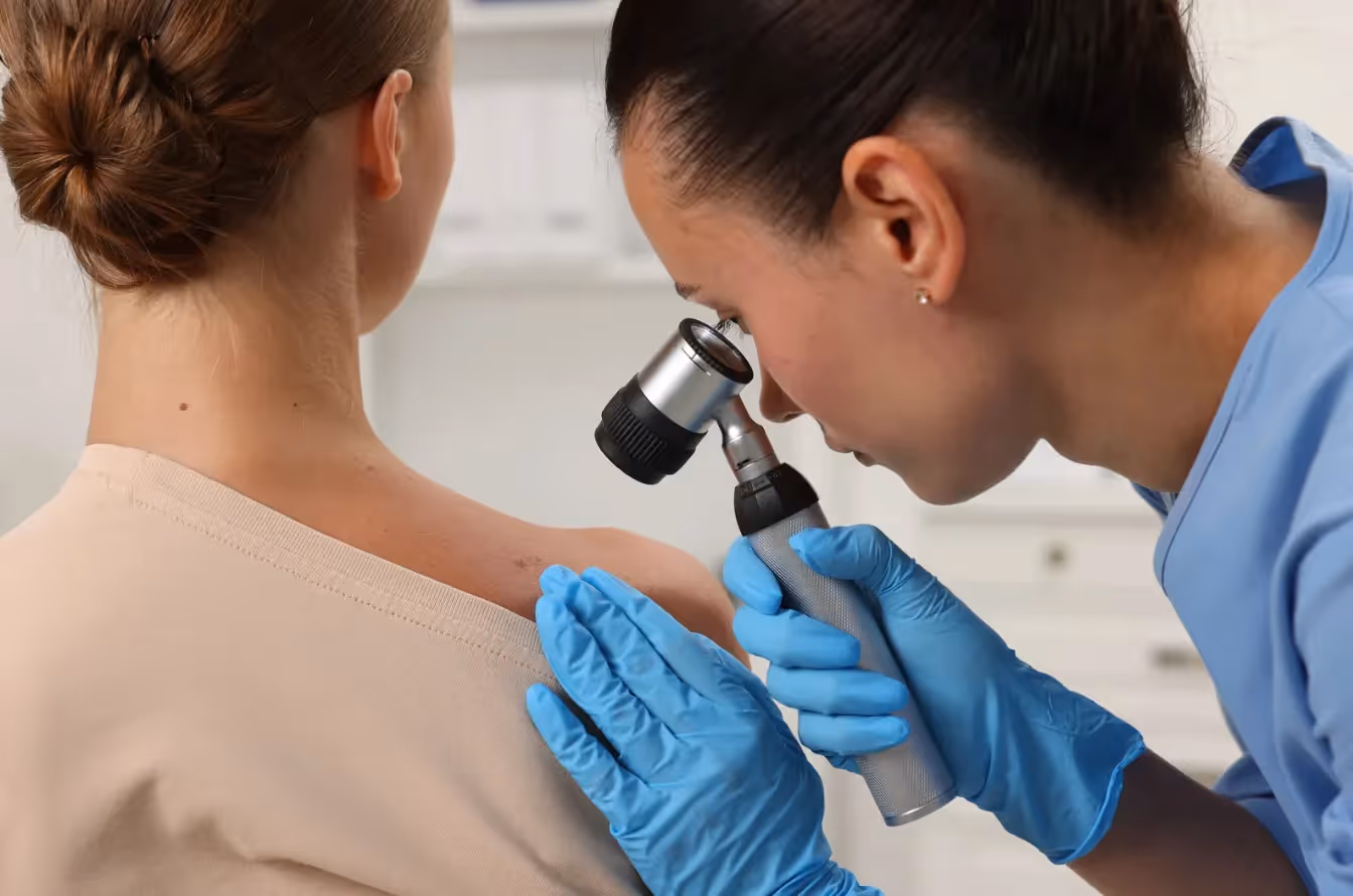
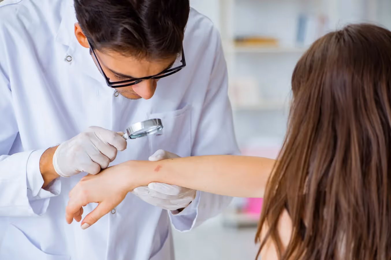
A pyogenic granuloma is benign, meaning it is not cancerous and not a sign of a serious underlying disease. However, heavy or repeated bleeding can be alarming and disruptive, and in some cases a lesion that bleeds frequently enough can cause minor anemia or become infected due to repeated wound exposure. The team at Hazany Derm recommends evaluation any time a skin lesion bleeds repeatedly or unexpectedly. Proper diagnosis is important because other lesions, including some that can be cancerous, may look similar to a pyogenic granuloma and require different management.
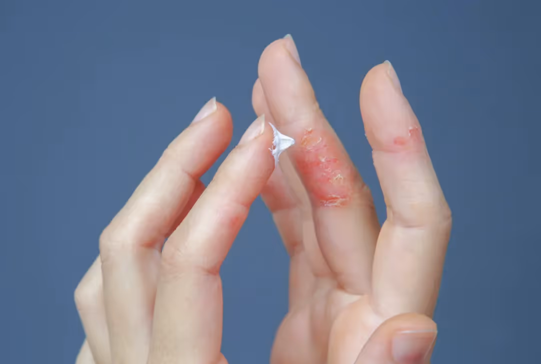
Can I treat a bleeding pyogenic granuloma at home?
While applying firm, steady pressure with a clean cloth can temporarily stop active bleeding from a pyogenic granuloma, home treatment does not address the underlying lesion. The fragile blood vessels remain intact, and the lesion will bleed again with the next contact or movement. Over-the-counter products and home cautery kits are generally ineffective and can cause scarring without achieving complete removal. Patients dealing with recurring bleeding from a skin growth are encouraged to schedule a consultation with a qualified dermatologist to get a proper assessment and discuss professional removal options.
Will a pyogenic granuloma go away on its own without treatment?
In most adults, pyogenic granulomas do not resolve spontaneously. Unlike some vascular lesions in infants that naturally regress over time, adult pyogenic granulomas typically persist and continue to bleed until they are treated. Pregnancy-related lesions sometimes shrink after delivery due to hormonal changes, but even those often require treatment if they persist. At the office of Dr. Salar Hazany, patients receive a thorough evaluation to determine the most appropriate course of action, with removal being the definitive solution for lesions that continue to bleed or cause cosmetic concern.
What are the treatment options for removing a pyogenic granuloma?
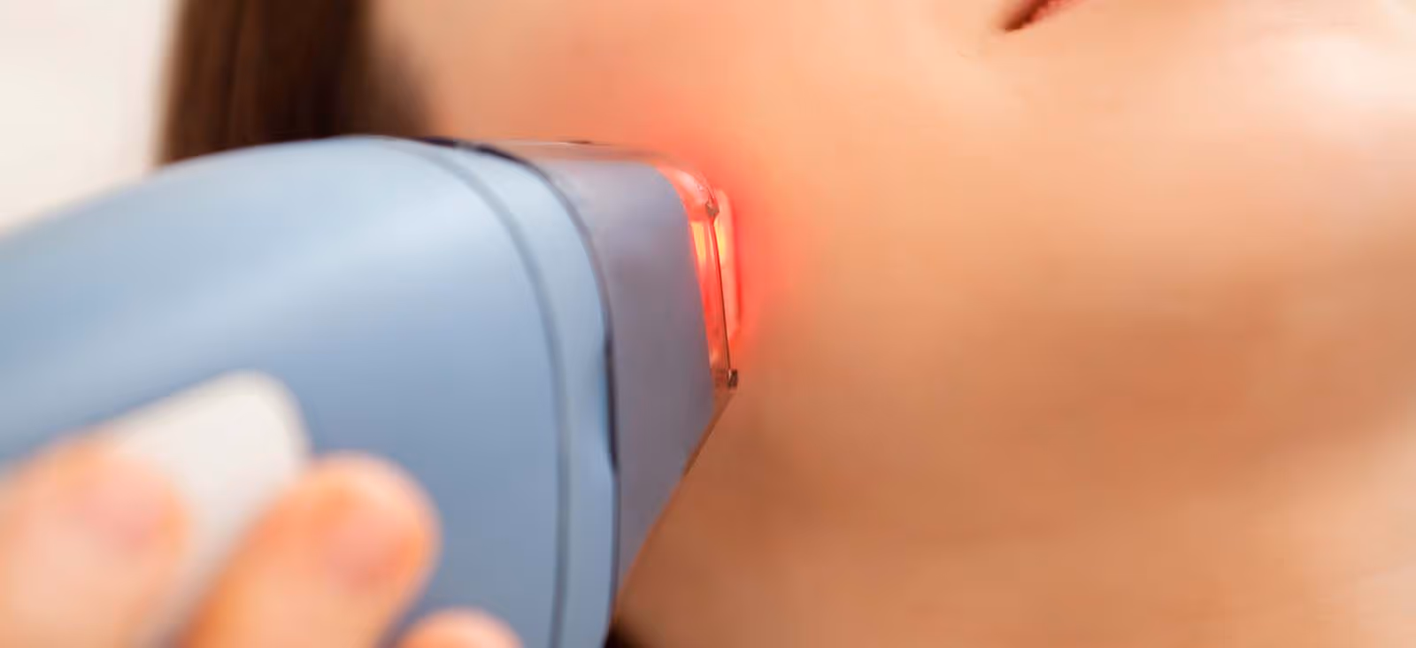
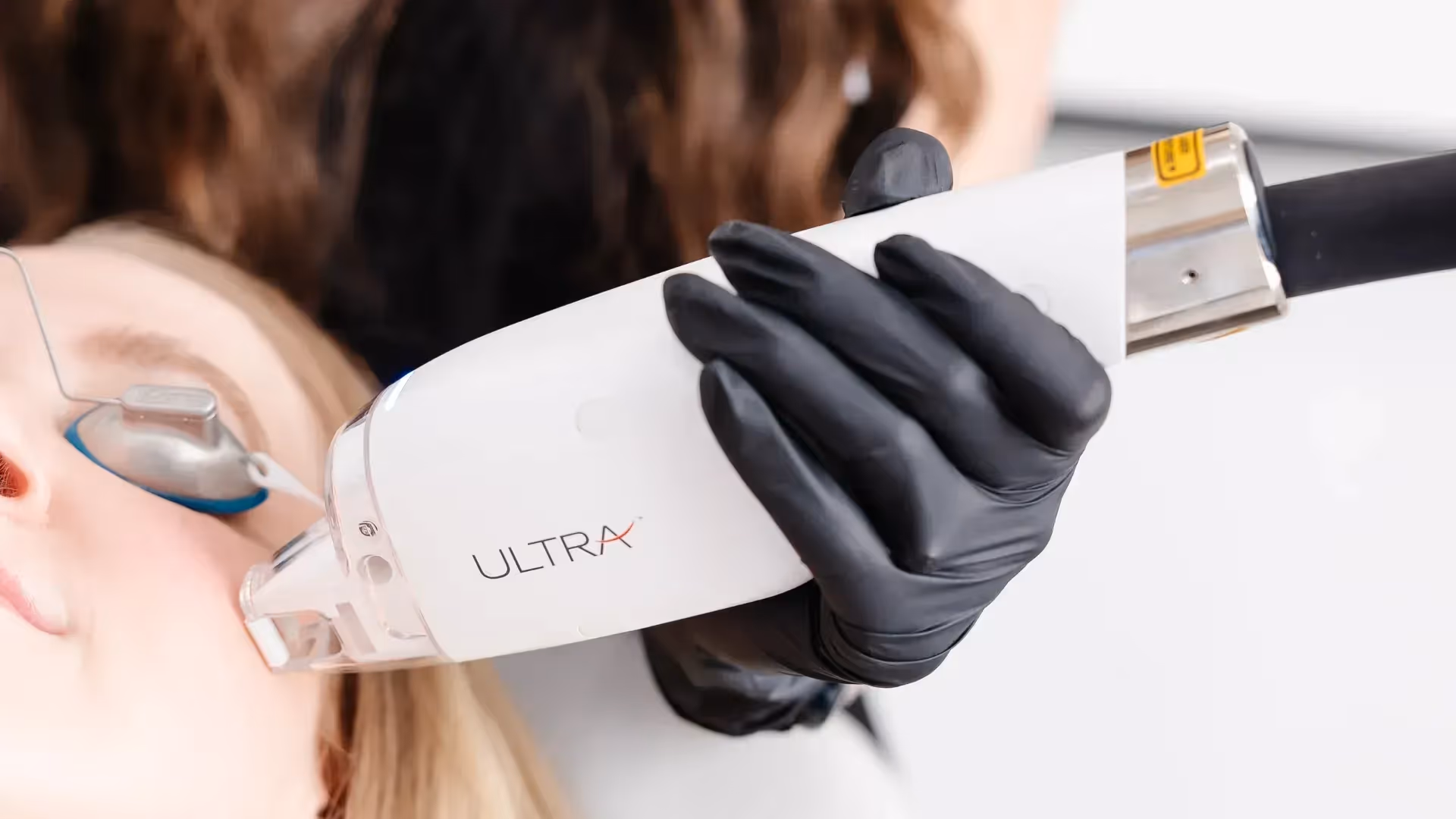
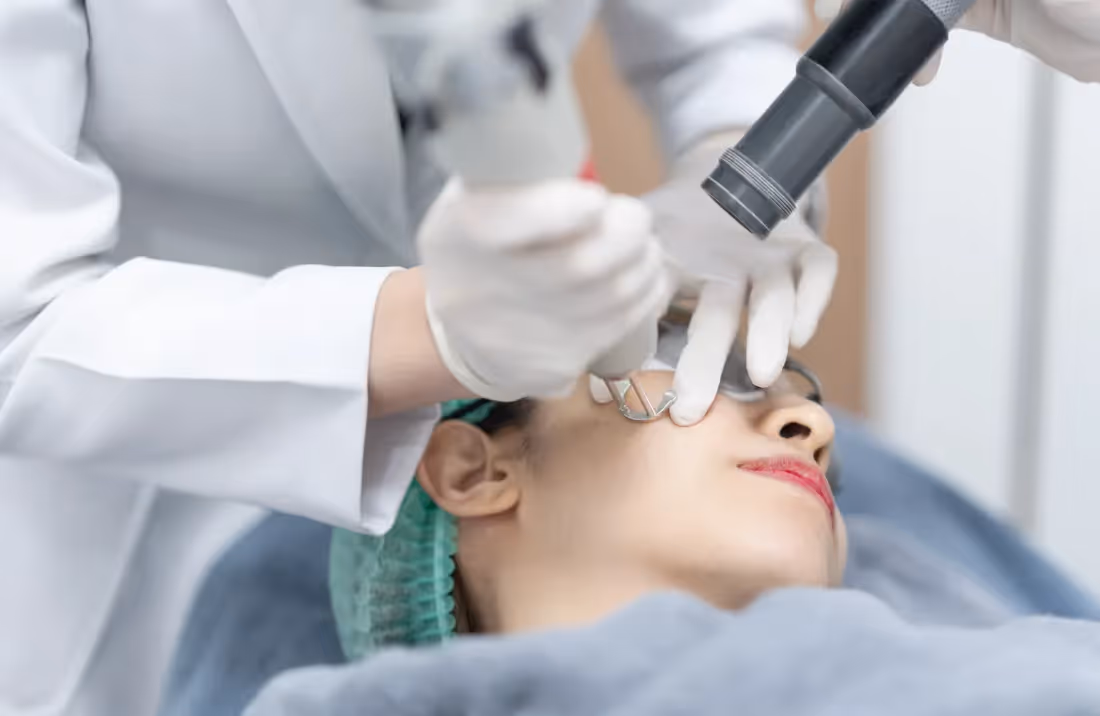
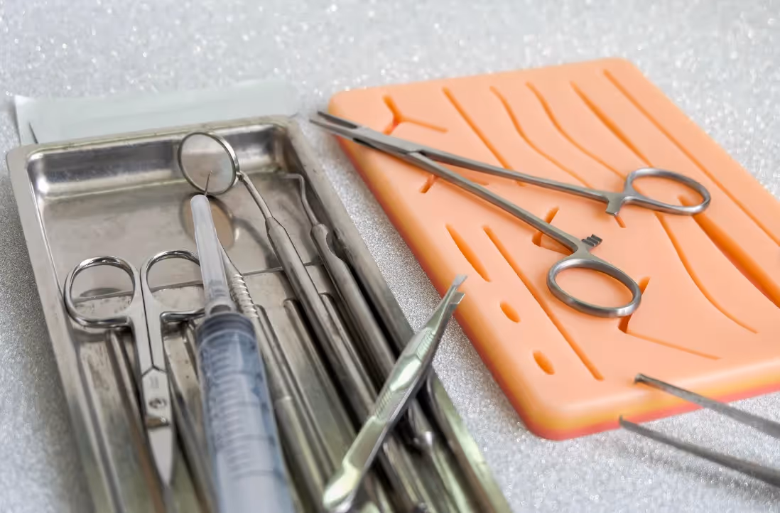
The most effective treatment for a pyogenic granuloma is professional removal, which can be accomplished through several methods depending on the lesion's size, location, and depth. Common approaches include shave excision with cauterization of the base, surgical excision with closure, laser treatment, or cryotherapy. Complete removal of the vascular base is critical to preventing recurrence. Hazany Derm offers precise, minimally invasive skin lesion removal tailored to each patient's anatomy and aesthetic goals, with particular expertise in treating lesions on sensitive areas like the nose, eyelid, and lip where both complete removal and cosmetic outcome matter greatly.
Why do pyogenic granulomas bleed more in some locations than others?
The location of a pyogenic granuloma significantly affects how often and how severely it bleeds, because different body areas vary in skin thickness, movement, and daily friction exposure. A lesion on the lip or eyelid is subject to constant motion and has very thin overlying skin, making bleeding nearly unavoidable during normal daily activities. A lesion on the back, by contrast, may bleed far less frequently. Dr. Hazany and his team at Hazany Derm take location into careful account when planning removal, using techniques specifically suited to the anatomical demands and cosmetic sensitivities of each individual treatment site.
Can a pyogenic granuloma come back after treatment?
Yes, pyogenic granulomas can recur after treatment, particularly if the base of the lesion is not completely addressed during removal. Partial treatment, such as surface cautery without removal of the deeper vascular lobules, often leads to regrowth at the same site within weeks to months. This is why the method and thoroughness of removal matter so much. The skin lesion removal approach used at Hazany Derm focuses on complete excision to minimize recurrence risk, giving patients the best chance of a permanent result without needing repeat procedures for the same lesion.
When should I see a doctor about a bleeding skin growth?
Any skin growth that bleeds repeatedly, grows rapidly, or changes in appearance deserves prompt professional evaluation. While pyogenic granulomas are benign, other skin lesions including amelanotic melanoma and other skin cancers can sometimes mimic their appearance. A trained dermatologist can examine the growth and, if necessary, send it for pathological analysis after removal to confirm the diagnosis. Consulting with a board-certified dermatologist is always the safest step when a skin lesion bleeds without clear cause. Patients in Los Angeles can visit Hazany Derm for expert evaluation and personalized treatment recommendations tailored to their specific lesion and skin type.