






%201.avif)



When Should a Mole Be Removed?



Moles should be removed when they show suspicious changes like asymmetry, irregular borders, color variation, or rapid growth. Professional evaluation at Hazany Derm ensures proper diagnosis and timely treatment of potentially dangerous lesions.
Moles are common skin growths that most people develop throughout their lives, typically appearing as small, dark spots on the skin. While the majority of moles are harmless and pose no health risks, some can develop into serious conditions that require immediate medical attention. Understanding when a mole should be removed is crucial for maintaining optimal skin health and preventing potential complications.
The decision to remove a mole involves careful consideration of various factors, including changes in appearance, location, and risk factors specific to each individual. Medical professionals use established criteria to evaluate moles and determine whether removal is necessary for diagnostic or cosmetic purposes. At Hazany Derm, patients receive comprehensive mole evaluations to ensure any concerning changes are identified and addressed promptly.
Early detection and intervention play vital roles in successful treatment outcomes when dealing with suspicious moles. Regular self-examinations combined with professional dermatological assessments create a comprehensive approach to mole monitoring. This proactive strategy helps identify problematic moles before they progress to more advanced stages that could compromise patient health and require more extensive treatment protocols.
Warning Signs That Indicate Mole Removal
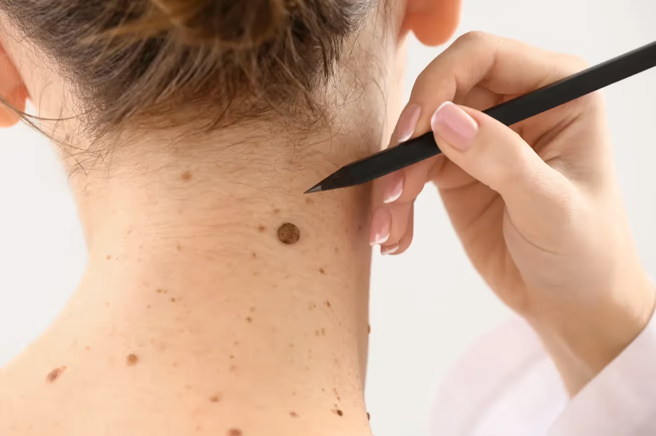
Recognizing the warning signs that indicate a mole should be removed requires understanding the ABCDE criteria used by dermatologists worldwide. These guidelines help both patients and medical professionals identify potentially dangerous moles that warrant immediate attention. Asymmetry, border irregularities, color variations, diameter changes, and evolution over time serve as key indicators that professional evaluation is necessary.
Changes in mole appearance often occur gradually, making them difficult to detect without systematic monitoring and documentation. Photographs taken at regular intervals can help track subtle modifications that might otherwise go unnoticed during casual observation. Dr. Salar Hazany emphasizes the importance of maintaining detailed records of mole characteristics to facilitate early detection of concerning changes.
The presence of multiple warning signs significantly increases the likelihood that a mole requires removal for proper histological examination. However, even a single suspicious characteristic should prompt immediate consultation with a qualified dermatologist. Professional assessment ensures accurate evaluation and appropriate treatment recommendations based on individual risk factors and clinical findings.
- Asymmetrical shape: When one half of the mole does not match the other half in size, shape, or appearance
- Irregular borders: Edges that are uneven, notched, scalloped, or poorly defined rather than smooth and regular
- Color variations: Multiple colors within a single mole, including different shades of brown, black, red, white, or blue
- Large diameter: Moles larger than 6 millimeters (about the size of a pencil eraser) or any mole that appears larger than others
Texture changes represent another important category of warning signs that often accompany visual modifications in problematic moles. Moles that become raised, develop a rough or scaly surface, or begin bleeding or oozing require immediate medical evaluation. These physical alterations frequently indicate cellular changes that could signify malignant transformation or other serious conditions requiring prompt intervention.
Itching, tenderness, or pain associated with a mole should never be ignored, as these symptoms often accompany dangerous cellular changes. Normal moles typically remain asymptomatic throughout their existence, making any discomfort a significant red flag for potential problems. Patients experiencing these symptoms should schedule dermatological consultations immediately to prevent potential complications and ensure appropriate treatment.
Medical Indications for Mole Removal
Certain medical conditions and risk factors create situations where mole removal becomes necessary regardless of apparent changes in the lesion's appearance. Individuals with family histories of melanoma or other skin cancers face elevated risks that may justify prophylactic removal of suspicious moles. Personal history of skin cancer, immunosuppression, or extensive sun exposure also contributes to increased risk profiles that influence treatment decisions.
Moles located in areas prone to friction or trauma often require removal to prevent irritation and potential complications. Clothing, jewelry, or regular shaving can cause repeated damage to moles, leading to inflammation, bleeding, or secondary infections. Hazany Derm specialists frequently recommend removing moles in high-friction areas to eliminate ongoing irritation and reduce the risk of complications from repeated trauma.
Atypical or dysplastic moles present unique challenges that often necessitate removal for accurate diagnosis and ongoing monitoring. These lesions exhibit characteristics that fall between normal moles and melanoma, requiring histological examination to determine their exact nature. Professional evaluation helps distinguish between benign atypical moles and those with malignant potential that require immediate intervention.
- Changing moles: Any mole that exhibits modifications in size, shape, color, texture, or sensation over time
- Bleeding or oozing: Moles that spontaneously bleed, discharge fluid, or develop open sores without trauma
- Family history: Strong genetic predisposition to melanoma or multiple atypical mole syndrome
- High-risk locations: Moles on the scalp, between fingers or toes, under nails, or in genital areas
Immunocompromised patients face particular challenges when dealing with potentially problematic moles, as their reduced immune function may accelerate malignant transformation. Organ transplant recipients, individuals receiving chemotherapy, or those with autoimmune conditions requiring immunosuppressive medications need more aggressive monitoring and earlier intervention. These patients benefit from regular professional evaluations and lower thresholds for mole removal recommendations.
The presence of multiple moles or atypical mole syndrome creates additional complexity in determining removal priorities and treatment strategies. Patients with numerous moles require systematic evaluation and risk stratification to identify the most concerning lesions. Dermatologists use specialized techniques including dermoscopy and digital mapping to monitor large numbers of moles efficiently and identify those requiring immediate attention or removal.
Cosmetic and Quality of Life Considerations
Beyond medical necessity, cosmetic concerns represent valid reasons for considering mole removal, particularly when lesions affect self-confidence or quality of life. Large, raised, or prominently located moles can cause significant psychological distress and social anxiety for some individuals. The team at Hazany Derm recognizes that cosmetic concerns are legitimate medical issues that deserve professional attention and appropriate treatment options.
Moles that interfere with daily activities, such as those caught by clothing or jewelry, create ongoing quality of life issues that justify removal. These practical concerns often coincide with cosmetic considerations, creating compelling cases for professional intervention. Removing problematic moles can eliminate daily frustrations and reduce the risk of trauma-related complications while improving overall patient satisfaction and comfort.
Professional mole removal ensures optimal cosmetic outcomes through proper technique selection and post-operative care protocols. Different removal methods produce varying cosmetic results, and experienced dermatologists can recommend the most appropriate approach for each individual situation. Advanced techniques minimize scarring and optimize healing to achieve the best possible aesthetic outcomes while ensuring complete lesion removal.
- Prominent facial moles: Lesions on highly visible areas that affect self-confidence and social interactions
- Large raised moles: Elevated lesions that catch on clothing, jewelry, or interfere with normal daily activities
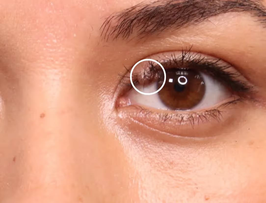
- Hair-bearing moles: Moles with coarse or dark hairs that create cosmetic concerns or grooming difficulties
- Multiple clustered moles: Groups of moles in visible areas that patients find aesthetically displeasing
The psychological impact of prominent moles should not be underestimated, as these lesions can significantly affect social interactions and self-perception. Studies demonstrate that cosmetic concerns related to visible skin lesions can lead to anxiety, depression, and social withdrawal in susceptible individuals. Professional removal of bothersome moles often produces dramatic improvements in self-confidence and overall quality of life that extend far beyond the cosmetic benefits.
Insurance coverage for mole removal varies depending on the specific indications and medical necessity of the procedure. Medically indicated removals typically receive coverage, while purely cosmetic procedures may require out-of-pocket payment. Patients should discuss insurance considerations with their dermatologist and insurance provider to understand coverage options and make informed decisions about treatment timing and approaches.
Frequently Asked Questions
What are the ABCDE warning signs that indicate a mole should be removed?
The ABCDE criteria represent the gold standard for identifying suspicious moles that require professional evaluation. A stands for Asymmetry, where one half of the mole doesn't match the other half. B represents Border irregularities, including uneven, notched, or poorly defined edges. C indicates Color variations, particularly multiple colors within a single lesion or unusual hues like red, white, or blue. D refers to Diameter, with moles larger than 6 millimeters being more concerning. E stands for Evolution, meaning any changes in size, shape, color, or texture over time. Dr. Salar Hazany emphasizes that even one of these characteristics warrants immediate professional assessment, as early detection significantly improves treatment outcomes and reduces complications.
How quickly should I see a dermatologist if I notice changes in a mole?
Any noticeable changes in a mole should prompt immediate consultation with a qualified dermatologist, ideally within one to two weeks of observation. Time is crucial when dealing with potentially malignant lesions, as early intervention dramatically improves treatment success rates and reduces the risk of metastasis. Rapid changes, bleeding, or sudden development of symptoms require urgent attention, sometimes within days rather than weeks. Hazany Derm prioritizes urgent mole evaluations and can accommodate patients with concerning changes through expedited scheduling. Don't adopt a "wait and see" approach with suspicious moles, as delays in diagnosis and treatment can have serious consequences. Professional evaluation provides peace of mind and ensures appropriate intervention when necessary.
Are there moles that should be removed even if they haven't changed?
Yes, certain moles warrant removal even without apparent changes, particularly those in high-risk categories or problematic locations. Moles with atypical or dysplastic characteristics may require prophylactic removal due to their increased malignant potential. Lesions located in areas prone to friction, such as under bra straps, waistbands, or razor areas, often need removal to prevent ongoing trauma and complications. Large congenital moles, especially those present since birth, carry elevated cancer risks that may justify early removal. Family history of melanoma or personal history of skin cancer can also influence removal decisions for otherwise stable moles. The team at Hazany Derm evaluates each patient's individual risk factors, mole characteristics, and personal circumstances to make appropriate removal recommendations that prioritize both medical safety and patient preferences.
What happens during a professional mole evaluation?
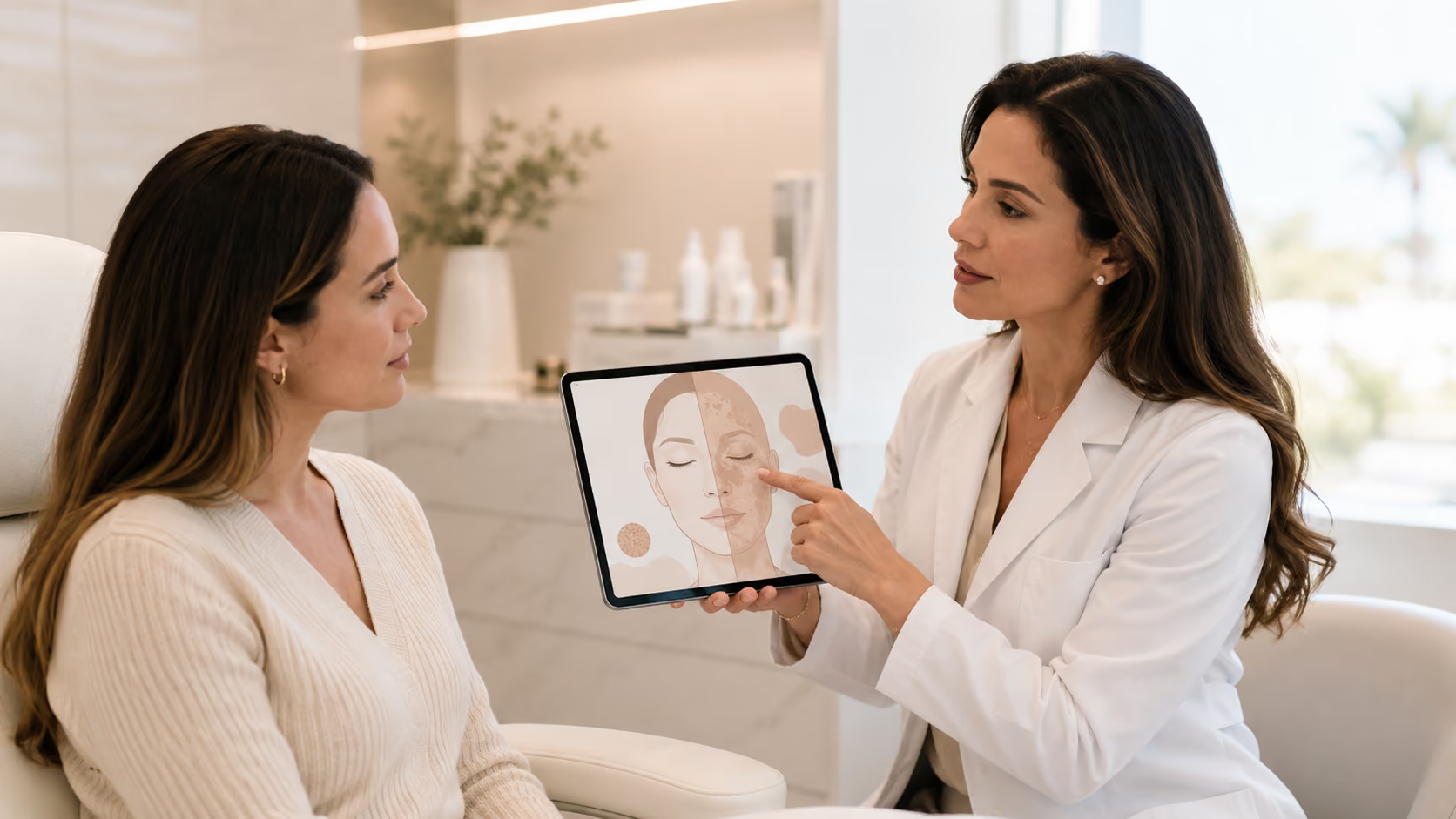
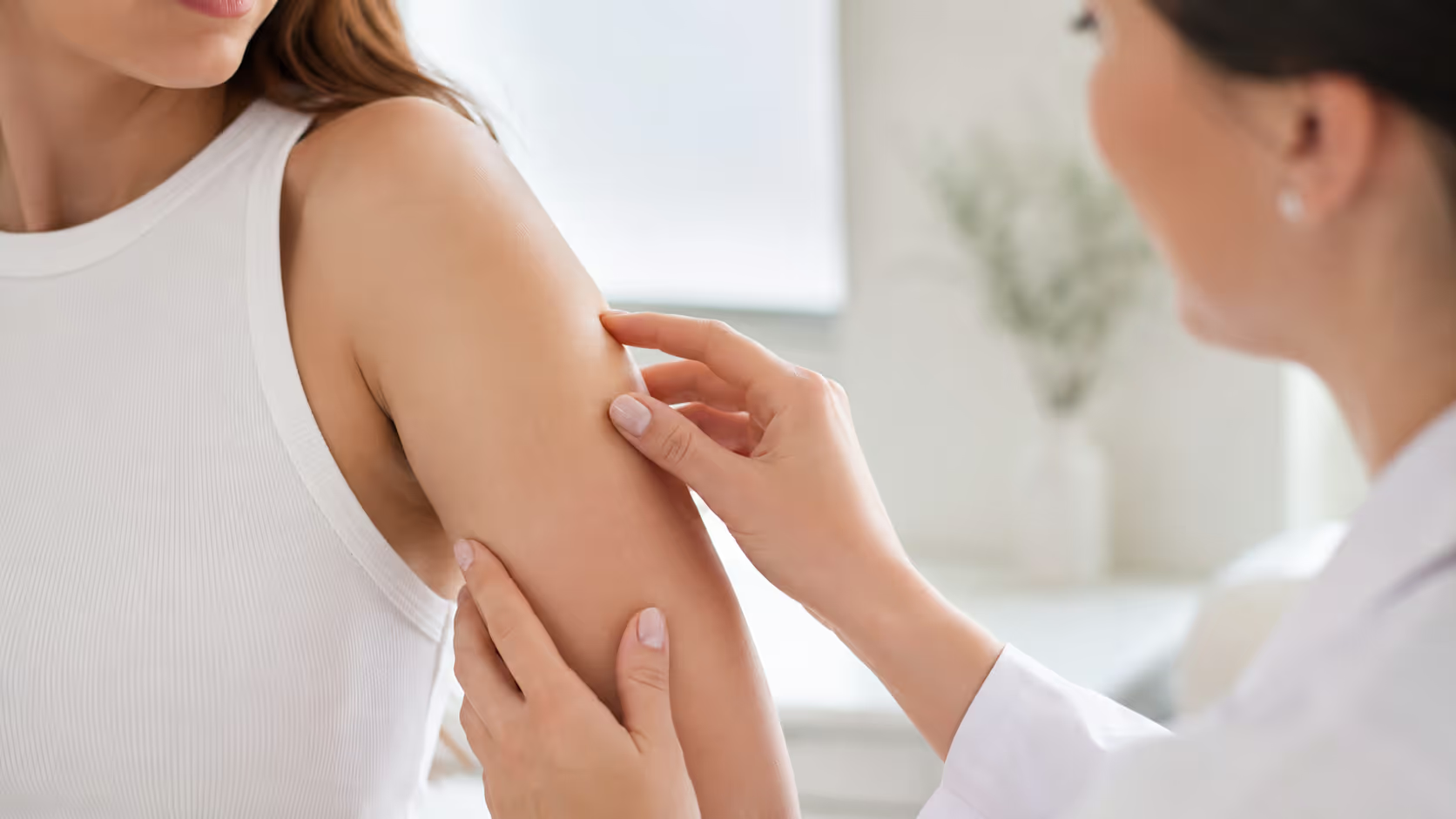
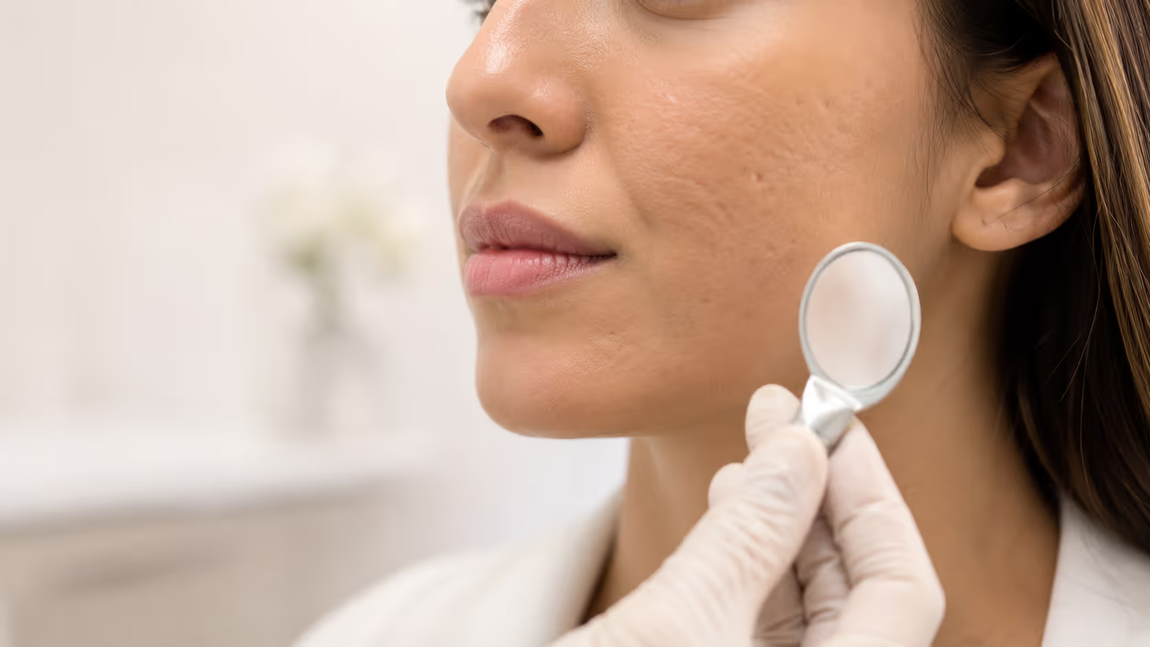
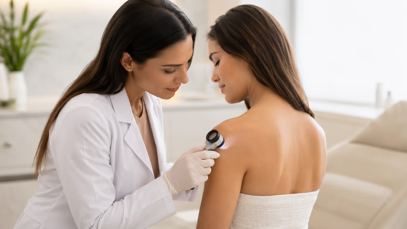
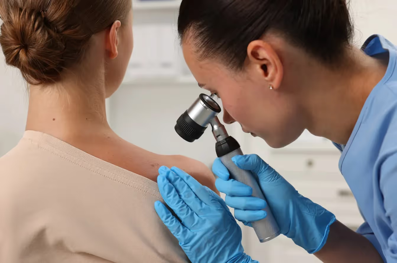
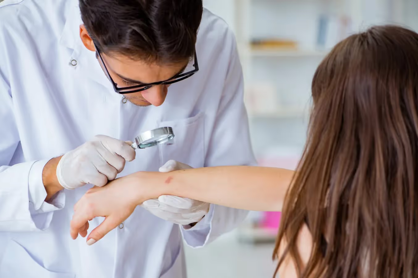
A comprehensive mole evaluation begins with a detailed medical history, including family history of skin cancer, personal sun exposure patterns, and any changes you've noticed in your moles. The dermatologist performs a thorough visual examination of your entire skin surface, often using specialized magnification tools called dermatoscopes to examine moles in greater detail. Digital photography may be used to document suspicious lesions for future comparison and monitoring purposes. Dr. Hazany also discusses your individual risk factors, lifestyle considerations, and any concerns you may have about specific moles. If suspicious characteristics are identified, the dermatologist will explain removal options, biopsy procedures, and expected outcomes. The evaluation concludes with personalized recommendations for ongoing monitoring, sun protection strategies, and follow-up scheduling based on your individual risk profile and examination findings.
What are the different methods for removing moles?
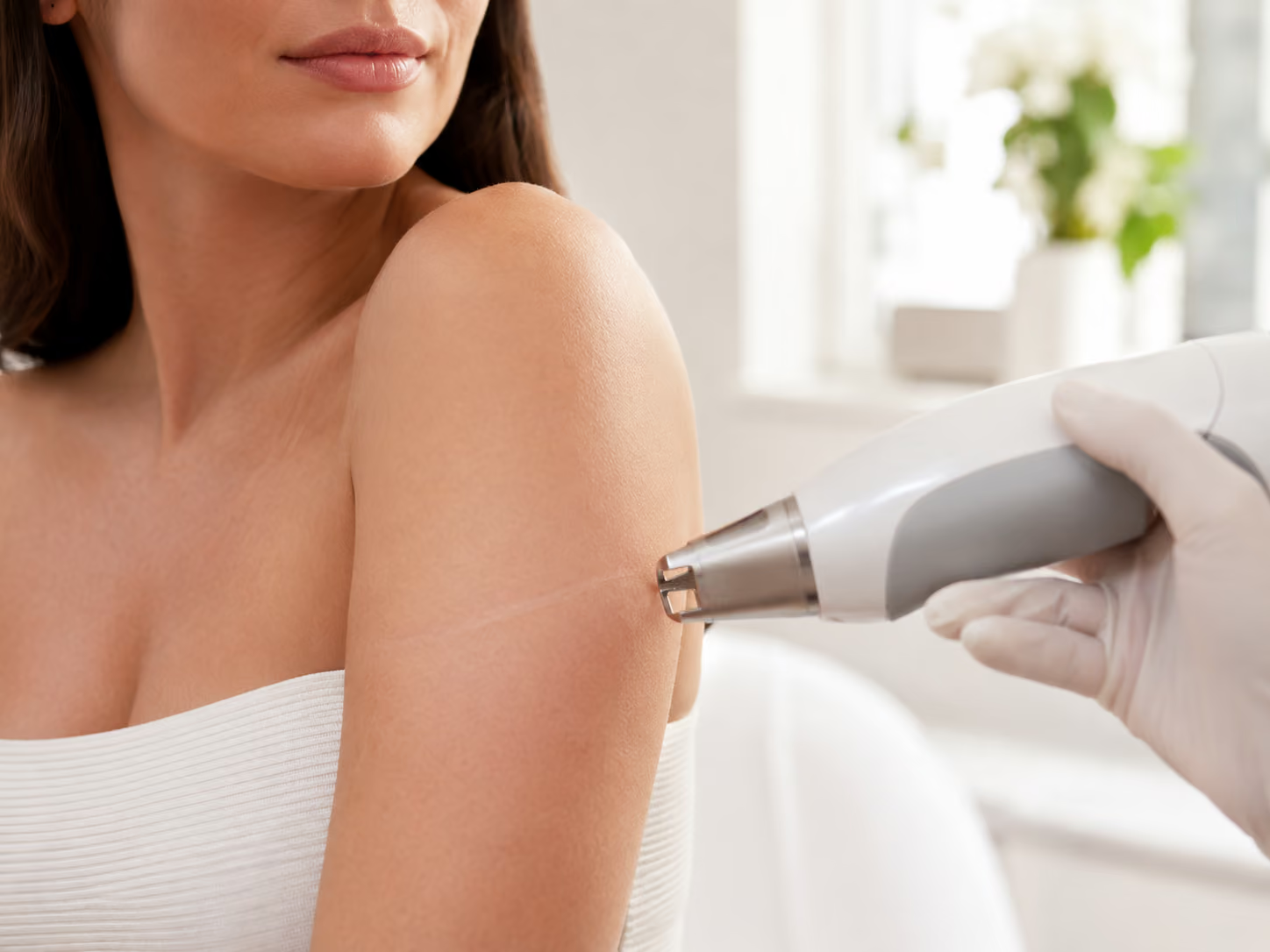
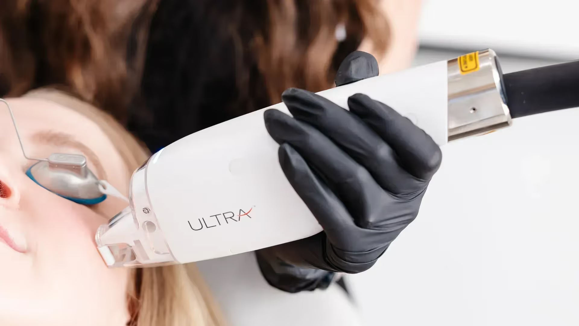
Several mole removal techniques are available, each suited to different types of lesions and patient circumstances. Shave excision involves using a sharp blade to remove raised moles at skin level, typically used for benign-appearing lesions that don't require deep tissue examination. Punch biopsy utilizes a circular cutting instrument to remove small, suspicious moles completely, providing tissue for histological analysis. Surgical excision involves cutting out the entire mole along with a margin of surrounding healthy tissue, often used for larger or more suspicious lesions. Laser removal may be appropriate for certain cosmetic cases, though it doesn't provide tissue for pathological examination. Hazany Derm specialists select the most appropriate removal method based on the mole's characteristics, location, suspected pathology, and desired cosmetic outcomes. The choice of technique significantly impacts healing time, scarring potential, and the ability to obtain diagnostic information.
Will mole removal leave a scar?
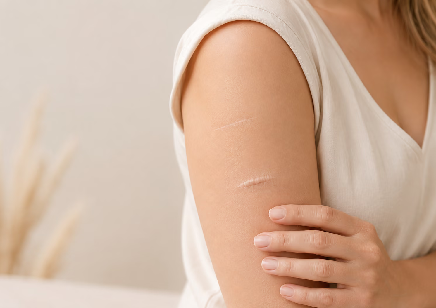
All mole removal procedures result in some degree of scarring, though the extent varies significantly based on the removal method, mole size and location, individual healing characteristics, and post-operative care. Shave excisions typically produce minimal, flat scars that often become nearly invisible over time. Surgical excisions create linear scars that, when properly closed and cared for, usually heal as thin, pale lines. Factors affecting scar appearance include the patient's age, skin type, genetic predisposition to scarring, and adherence to post-operative care instructions. Dr. Salar Hazany employs advanced surgical techniques and provides comprehensive aftercare guidance to optimize healing and minimize scarring. Most patients find that any residual scarring is far preferable to living with a concerning or bothersome mole. Proper wound care, sun protection during healing, and follow-up appointments help ensure the best possible cosmetic outcomes.
Is mole removal covered by insurance?
Insurance coverage for mole removal depends primarily on the medical necessity of the procedure and the specific characteristics of your insurance plan. Medically indicated removals, such as those for suspicious or changing moles, atypical lesions, or moles causing physical problems, typically receive full or partial coverage. Purely cosmetic removals for aesthetic reasons alone usually require out-of-pocket payment, though some plans may offer partial coverage. Pre-authorization may be required for certain procedures, and documentation of medical necessity often needs to be provided to insurance companies. The Hazany Derm clinic works closely with patients and insurance providers to maximize coverage opportunities and provide clear cost estimates before procedures. Patients should contact their insurance provider directly to understand their specific coverage details, deductibles, and any pre-authorization requirements that may apply to their situation.
How should I monitor my moles between dermatologist visits?
Regular self-examination represents a crucial component of comprehensive mole monitoring and early skin cancer detection. Perform monthly self-checks in good lighting using a full-length mirror and hand mirror to examine all skin surfaces, including often-overlooked areas like between toes, under arms, and on the scalp. Take photographs of concerning or atypical moles to track changes over time, as subtle modifications can be difficult to detect through memory alone. Use the ABCDE criteria to systematically evaluate each mole, noting any asymmetry, border irregularities, color variations, large diameter, or evolution over time. Keep a written log of new moles or changes in existing ones, including dates and descriptions of modifications. Schedule professional evaluations immediately if you notice any suspicious changes, rather than waiting for your next routine appointment. The experts at Hazany Derm provide detailed self-examination instructions and welcome patient questions about any concerning observations between scheduled visits.