






%201.avif)



What Should I Do if I Find a Suspicious Mole?



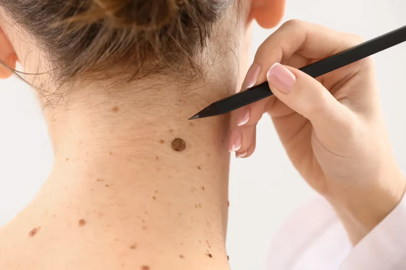
If you find a suspicious mole, schedule an immediate dermatological evaluation. Document changes with photos, avoid self-diagnosis, and seek professional assessment. The experienced team at Hazany Derm provides comprehensive mole evaluations and expert skin cancer screening services.
Discovering a suspicious mole can be alarming, but taking swift and appropriate action is crucial for your health and peace of mind. Moles are common skin growths that most people develop throughout their lives, but certain changes in appearance, texture, or behavior warrant immediate professional attention. Understanding what makes a mole suspicious and knowing the proper steps to take can potentially save your life.
The key to successful treatment of any skin abnormality lies in early detection and prompt medical evaluation. While many mole changes are benign, some may indicate the presence of melanoma or other forms of skin cancer that require immediate intervention. The difference between a routine mole check and an urgent dermatological consultation often depends on recognizing specific warning signs and acting decisively.
At Hazany Derm, we emphasize the importance of patient education and proactive skin health management. Dr. Salar Hazany and his team regularly encounter patients who have discovered concerning changes in their moles, and the outcomes are consistently better when individuals seek professional evaluation quickly rather than waiting or attempting self-diagnosis. Taking the right steps immediately after noticing suspicious changes can make all the difference in your treatment outcomes and long-term health.
Immediate Steps to Take When You Notice a Suspicious Mole
The moment you identify a potentially problematic mole, your first priority should be scheduling a dermatological appointment as soon as possible. Time is often a critical factor in skin cancer treatment, and delays can allow potentially dangerous conditions to progress unnecessarily. Most dermatology practices, including Hazany Derm, prioritize urgent consultations for suspicious skin lesions and can often accommodate same-day or next-day appointments for concerning cases.
While waiting for your appointment, document the suspicious mole thoroughly by taking high-quality photographs from multiple angles. Use good lighting and include a reference object like a coin or ruler to show the mole's size accurately. Take photos of the mole from different distances to capture both the overall appearance and fine details that might be relevant to your dermatologist's evaluation.
Resist the urge to manipulate, pick at, or attempt to remove the suspicious mole yourself, as this can interfere with accurate diagnosis and potentially cause complications. Home remedies, over-the-counter treatments, and DIY removal attempts can alter the mole's appearance, making it more difficult for your dermatologist to assess the original characteristics. Additionally, improper handling can lead to infection, scarring, or incomplete removal of potentially dangerous tissue.
- Document thoroughly: Take clear, well-lit photographs from multiple angles with size references
- Schedule immediately: Contact your dermatologist for the earliest possible appointment
- Avoid manipulation: Do not pick, scratch, or attempt home treatments on the suspicious area
- Monitor changes: Keep track of any additional changes while waiting for your appointment
After taking initial documentation photos, continue monitoring the mole for any additional changes while you wait for your dermatological appointment. Note any variations in size, color, texture, or symptoms like itching, bleeding, or pain, as this information will be valuable during your consultation. Keep a simple log or diary of observations, including dates and descriptions of any changes you notice.
Share your concerns and findings with family members or close friends who can provide support and help you remember important details about the mole's evolution. Having another person aware of the situation can be helpful for emotional support and practical assistance, such as transportation to appointments or help with follow-up care if needed. At Hazany Derm, we often find that patients who have strong support systems tend to follow through more consistently with recommended treatments and monitoring protocols.
Understanding the ABCDE Warning Signs
The ABCDE criteria represent the gold standard for identifying potentially dangerous moles and are widely used by dermatologists worldwide, including the expert team at Hazany Derm. These five warning signs provide a systematic approach to mole evaluation that both patients and healthcare providers can use to identify lesions requiring professional attention. Understanding these criteria empowers you to conduct regular self-examinations and recognize when changes warrant immediate medical evaluation.
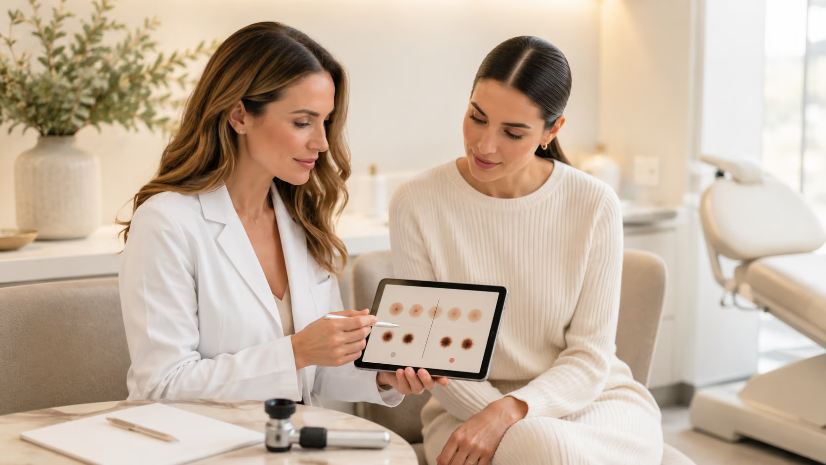
"A" stands for Asymmetry, which means that one half of the mole does not match the other half in shape, color, or texture. Normal, benign moles are typically symmetrical, meaning you could draw a line through the center and both halves would look essentially the same. Asymmetrical moles often indicate abnormal cell growth patterns that may suggest malignant transformation and require professional evaluation.
"B" represents Border irregularity, referring to moles with edges that are uneven, notched, scalloped, or poorly defined. Healthy moles usually have smooth, even borders that create a clear distinction between the mole and surrounding skin. Irregular borders can indicate that the cells are growing in an uncontrolled pattern, which is characteristic of malignant lesions and requires immediate dermatological assessment.
- Asymmetry: One half of the mole doesn't match the other half in appearance
- Border irregularity: Edges are uneven, notched, or poorly defined rather than smooth
- Color variation: Multiple colors within the same mole or significant color changes
- Diameter increase: Growth beyond 6 millimeters or notable size increases over time
"C" indicates Color variation or changes, including multiple colors within a single mole or significant alterations in existing coloration. Normal moles are typically uniform in color, whether brown, black, or tan, while suspicious moles may display multiple shades or develop new colors like red, white, or blue. Any dramatic color changes in existing moles, such as darkening, lightening, or the appearance of new hues, should prompt immediate professional evaluation.
"D" and "E" represent Diameter and Evolution respectively, with diameter referring to moles larger than 6 millimeters (about the size of a pencil eraser) and evolution describing any changes in size, shape, color, or symptoms over time. While not all large moles are dangerous, size increases or moles that develop symptoms like itching, bleeding, or crusting require professional assessment. Dr. Salar Hazany emphasizes that the "E" for evolution is often the most important factor, as any changing mole deserves immediate attention regardless of whether it meets other ABCDE criteria.
Professional Evaluation and Diagnostic Process
When you arrive for your dermatological consultation at Hazany Derm, the evaluation process begins with a comprehensive review of your medical history, including previous skin cancers, family history of melanoma, and any medications or conditions that might affect your skin health. Dr. Salar Hazany and his team will also discuss your sun exposure history, use of tanning beds, and any previous concerning moles or skin lesions. This background information helps establish your risk level and informs the approach to your current concern.
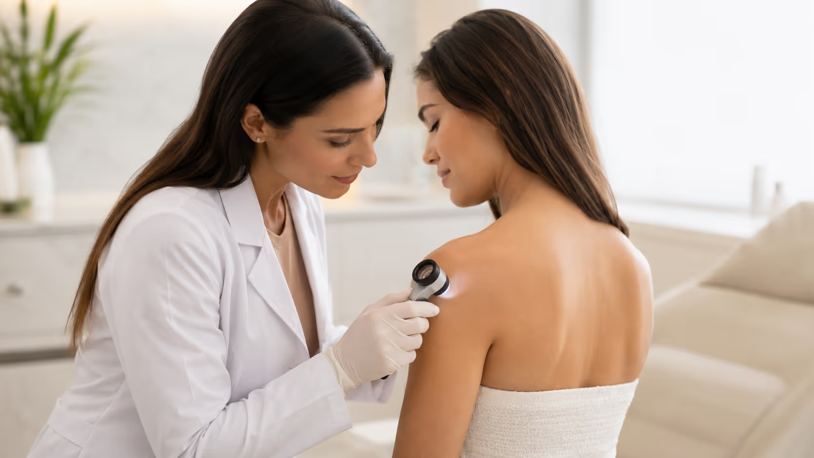
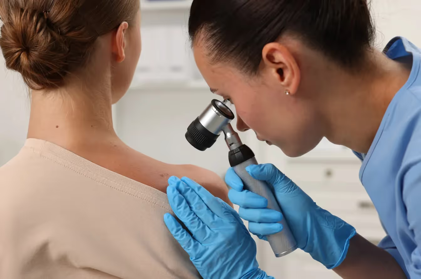
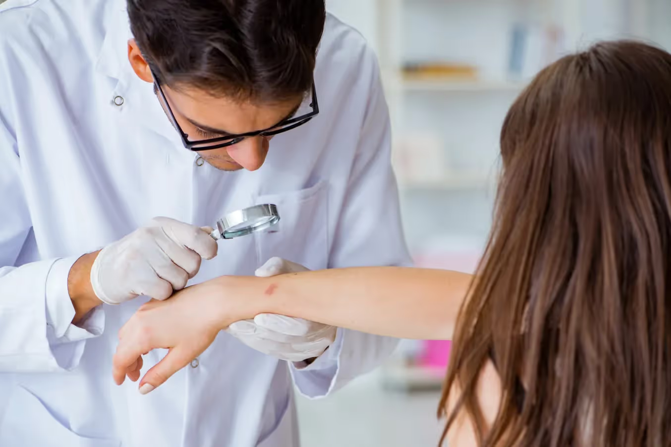
The physical examination involves a detailed inspection of the suspicious mole using specialized equipment, including dermatoscopy, which provides magnified visualization of skin structures not visible to the naked eye. This non-invasive technique allows dermatologists to examine the mole's internal patterns, blood vessel arrangements, and pigment distribution with remarkable detail. The dermatoscopic evaluation often provides sufficient information to determine whether a mole is likely benign or requires tissue sampling for definitive diagnosis.
Depending on the findings during the clinical examination, your dermatologist may recommend a biopsy to obtain tissue for microscopic analysis. There are several biopsy techniques available, including shave biopsies, punch biopsies, and excisional biopsies, with the choice depending on the mole's characteristics, location, and suspected diagnosis. The biopsy procedure is typically performed in the office under local anesthesia and provides definitive information about the nature of the suspicious lesion.
- Medical history review: Comprehensive discussion of risk factors and previous skin issues
- Dermatoscopic examination: Magnified visualization using specialized optical equipment
- Clinical photography: Professional documentation for monitoring and treatment planning
- Biopsy recommendation: Tissue sampling when clinical examination suggests potential malignancy
Following the biopsy procedure, tissue samples are sent to specialized dermatopathologists who analyze the cellular structure and characteristics under microscopic examination. Results are typically available within 5-7 business days, though urgent cases may receive expedited processing when clinically indicated. The pathology report provides detailed information about the type of cells present, their growth patterns, and whether any malignant characteristics are identified.
At Hazany Derm, patients receive clear, comprehensive explanations of their biopsy results along with detailed discussions of treatment options and follow-up recommendations. Whether the results show benign changes, precancerous conditions, or malignant findings, Dr. Salar Hazany ensures that patients understand their diagnosis and feel confident about their treatment plan. The team provides ongoing support throughout the diagnostic process and coordinates any additional treatments or referrals that may be necessary based on the findings.
Frequently Asked Questions
What are the most concerning signs that indicate a mole needs immediate medical attention?
The most alarming signs requiring immediate evaluation include rapid changes in size, shape, or color, bleeding or oozing, persistent itching or pain, and irregular borders or asymmetry. Dr. Salar Hazany emphasizes that any mole displaying multiple colors, particularly red, white, or blue areas within a brown or black lesion, warrants urgent assessment. Additionally, new moles appearing after age 30, especially those that grow quickly or look different from your other moles, should be evaluated promptly. The key is recognizing that any changing mole is potentially dangerous and requires professional evaluation rather than watchful waiting.
How quickly should I schedule an appointment after noticing a suspicious mole?
You should contact a dermatologist within 24-48 hours of identifying suspicious changes in a mole, as early detection significantly improves treatment outcomes for potential skin cancers. Hazany Derm prioritizes urgent consultations for concerning skin lesions and often accommodates same-day appointments when patients report alarming symptoms like bleeding, rapid growth, or dramatic color changes. While not every suspicious mole represents an emergency, the potential consequences of delayed diagnosis make prompt evaluation essential. Most dermatology practices understand the anxiety associated with suspicious moles and work to minimize wait times for these concerning cases.
Can I monitor a suspicious mole at home instead of seeing a doctor immediately?
Home monitoring should never replace professional evaluation when you've identified a suspicious mole, as self-diagnosis can lead to dangerous delays in treatment. The team at Hazany Derm strongly advises against the wait-and-see approach for any mole that meets ABCDE criteria or has undergone recent changes. While regular self-examinations are important for ongoing skin health, suspicious findings require dermatological expertise and potentially specialized diagnostic equipment like dermatoscopy. Professional evaluation can often provide immediate reassurance for benign changes or identify serious conditions that require prompt treatment, making the investment in professional assessment invaluable for your long-term health.
What should I do to prepare for my dermatology appointment?
Preparation involves documenting the suspicious mole with clear photographs, noting when changes first appeared, and compiling relevant medical history including previous skin cancers and family history. Dr. Hazany recommends removing nail polish and makeup to allow thorough skin examination, and wearing comfortable clothing that provides easy access to the concerning area. Prepare a list of current medications, previous skin treatments, and any symptoms associated with the mole such as itching, bleeding, or pain. Additionally, consider bringing a trusted friend or family member for support and to help remember important information discussed during the consultation.
What types of biopsies might be recommended for a suspicious mole?
Several biopsy techniques may be appropriate depending on the mole's characteristics, including shave biopsies that remove the raised portion, punch biopsies that take a deeper cylindrical sample, and excisional biopsies that remove the entire lesion with surrounding tissue. Hazany Derm specialists select the most appropriate technique based on the suspected diagnosis, lesion size and location, and cosmetic considerations. Shave biopsies are common for raised lesions, while flat or deeply pigmented moles may require punch or excisional techniques. All biopsy procedures are performed under local anesthesia in the office setting, with most patients experiencing minimal discomfort and returning to normal activities within 24-48 hours.
How accurate are dermatological evaluations in detecting skin cancer?
Dermatological evaluations by experienced professionals like Dr. Salar Hazany have high accuracy rates, particularly when combined with dermatoscopy and appropriate biopsy techniques when indicated. Clinical examination by trained dermatologists can identify suspicious lesions with approximately 85-90% accuracy, while dermatoscopy increases diagnostic precision significantly. However, definitive diagnosis often requires histopathological examination of biopsy samples, which provides near 100% accuracy in determining the exact nature of suspicious lesions. The combination of clinical expertise, advanced diagnostic tools, and laboratory analysis creates a comprehensive evaluation system that maximizes early detection while minimizing unnecessary procedures for benign conditions.
What happens if my biopsy results show skin cancer?
If biopsy results confirm skin cancer, the Hazany Derm clinic will immediately develop a comprehensive treatment plan tailored to the specific type, stage, and location of the cancer. Treatment options may include surgical excision, Mohs surgery for sensitive areas, topical therapies for superficial cancers, or referral to oncology specialists for advanced cases. The clinic coordinates all aspects of care, from initial treatment through follow-up monitoring and preventive education. Most skin cancers, particularly those detected early, have excellent cure rates with appropriate treatment. The team provides detailed explanations of treatment options, expected outcomes, and ongoing surveillance protocols to ensure optimal results and early detection of any future concerns.
How can I prevent future suspicious moles from developing?
Prevention strategies include consistent use of broad-spectrum sunscreen with SPF 30 or higher, seeking shade during peak sun hours, wearing protective clothing and wide-brimmed hats, and avoiding tanning beds entirely. The experts at Hazany Derm emphasize that cumulative sun exposure throughout life contributes to skin cancer risk, making daily sun protection essential regardless of age or previous sun damage. Regular self-examinations monthly and professional skin checks annually or as recommended help identify new or changing lesions early. Additionally, maintaining awareness of your personal and family history of skin cancer, understanding your individual risk factors, and promptly addressing any suspicious changes contribute significantly to long-term skin health and cancer prevention.